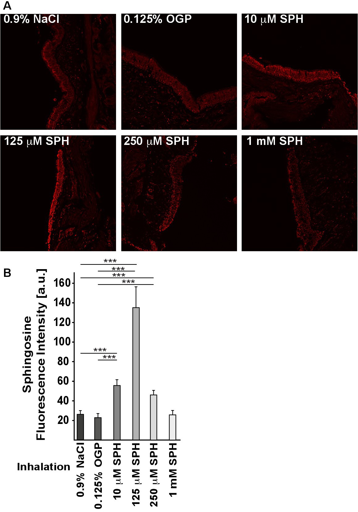
Fig. 1. Histological studies demonstrate an accumulation of sphingosine in bronchial epithelial cells after inhalation. Mini pigs were inhaled with sphingosine at the indicated concentration, with 0.9% NaCl alone or with 0.125% octylglucopyranoside (OGP) as controls. The pigs were subjected to bronchoscopy 17 hrs after the last sphingosine inhalation, biopsies were fixed in paraformaldehyde, embedded in paraffin and sectioned. Sections were stained with Cy3-coupled anti-sphingosine antibodies. Shown are representative immune stainings (A) and the quantitative analysis of the fluorescence intensity of a total of 1200 cells per group (B). Given is the mean ± SD, *p<0.05, **p<0.01, ***p<0.001, ANOVA.